
Study & Master
Support Pack | Grade 11
CAPS
Life Sciences
Life processes in plants and animals
This support pack for the Life processes in plants and animals
strand of the Life Sciences Grade 11 CAPS curriculum
provides revision summaries on the topic to help prepare for the
examinations. Learners can work through these individually at
home or these could form the basis of a catch-up class or online
lesson. You have permission to print or photocopy this document
or distribute it electronically via email or WhatsApp.
Cambridge University Press Africa
is a proudly South African publisher.
For more information on our Study & Master CAPS-approved
textbooks and valuable resource materials, visit
www.cambridge.org
Brighter thinking | Better learning
www.cambridge.org
Skip to contentStudy guide 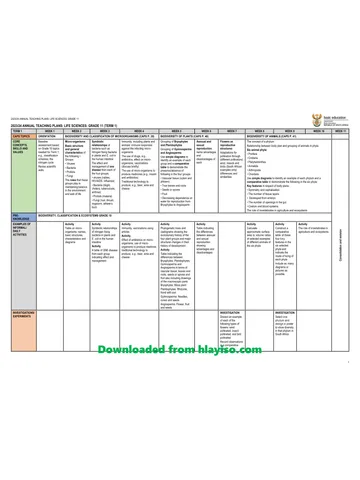
View all
3 . G11 LifeSciences DigitalSupport web Life Processes in Plants and Animals
Grade 11 Life Sciences study guide, 19 pages. Read online or get the free PDF download.
Publisher: Study & Master
Life Sciences · Grade 11. Study guide, 19 pages. Read online or download the PDF.
- Subject
- Life Sciences
- Grade
- Grade 11
- Document type
- Study guide
- Publisher
- Study & Master
- Curriculum
- CAPS
- Pages
- 19
- File size
- 1.2 MB
Loading document…
Loading document…
1 of 19
Document textSearch extracted text and jump to a page.
UNIT 1: Photosynthesis
formation of food storage of
in chloroplasts manufactured food
temperature
carbon dioxide
trapping and release of
storing energy oxygen
improving crop yields
in greenhouses by CO2
light enrichment, optimum
intake of raw light and
materials dark phases light and temperature
Photosynthesis:
Rate of photosynthesis
process and importance
Role of ATP as an important
Photosynthesis energy carrier in the cells
UNIT 1 Photosynthesis
• Photosynthesis is a process in which enzymes are used to build up complex
products from simple substrates – a biosynthetic or anabolic (building-up)
process.
• Photosynthesis takes place in special organelles in the cell called
chloroplasts.
• These equations describe the process of photosynthesis:
chlorophyll
Carbon dioxide + water + light → glucose + oxygen
enzymes
This equation can also be illustrated by symbols as follows:
chlorophyll
CO2 + H2O + light → C6H12O6 + O2
enzymes
• Photosynthesis requires light (radiant energy), water, carbon dioxide,
chlorophyll and enzymes, and produces oxygen and glucose.
• Photosynthesis takes place in the green parts of the plant, mainly in the
leaves, in organelles called chloroplasts.
• Chloroplasts contain the pigment chlorophyll, which traps light energy.
The importance of photosynthesis
• Photosynthesis is important because the process releases oxygen into the
atmosphere, uses carbon dioxide and traps energy for food production.
Factors affecting the rate of photosynthesis
• The rate of photosynthesis is determined by environmental conditions such
as temperature, light intensity and carbon dioxide concentration.
• Food producers such as farmers use their understanding of photosynthesis to
improve the growth of crops in greenhouses.
The role of adenosine triphosphate (ATP)
• ATP is the major energy carrier of the cell, providing the energy for most of
the energy-consuming activities of the cell.
UNIT 2: Animal nutrition (mammals)
structure and hepatic portal
importance of villi system
carbohydrases,
incorporation of
proteases, lipases role of the liver
small intestine glucose and amino
acids into cells
chemical digestion:
diabetes
enzymes
peristalsis
absorption assimilation
hormonal control of
mechanical or blood sugar levels
chewing physical digestion
Homeostatic control
types and functions of
different types of teeth Nutritional processes
and significance of
relationship between tooth decay
food intake, energy, related to
functions of different parts growth and health diet
requirements
Animal nutrition
Human nutrition
(mammals) malnutrition
different
diets
structure of alimentary dietary
canal and associated organs supplements
Dentition
differences in terms of Alcohol and drug abuse
nutritional requirements
and energy relationships herbivores
dangers associated
omnivores carnivores effects with their misuse
UNIT 2 Animal nutrition (mammals)
Herbivores, carnivores and omnivores
• Herbivores, carnivores and omnivores have different dentition because of
their different diets and lifestyles.
Human nutrition
• Humans have a two-ended (two-opening) digestive tract or alimentary canal.
The digestive system can be divided into a tubular alimentary canal and
accessory organs.
• Each section of the human digestive tract is very specialised; each performs
its own function.
nasal cavity
palate
tongue
bolus of food
salivary duct
epiglottis
salivary gland
trachea
oesophagus
diaphragm
liver
sphincter muscle
gall bladder
bile duct stomach
sphincter
muscle
pancreas
pancreatic duct
duodenum
small intestine
ileum
caecum
colon
appendix large intestine
rectum
anus
Figure 1 The human digestive system
The processes that are involved in the movement of food
through the digestive system
• The following processes take place during digestion in humans:
• Ingestion: the intake of food through the mouth
• Digestion: the mechanical and chemical breakdown of food
• Absorption: the diffusion of simple food molecules through the
intestinal wall into the blood
• Assimilation: the incorporation of nutrients such as amino acids and
glucose into the cells
• Egestion/defecation: the removal of undigested and unabsorbed waste
from the body through the anus in the form of faeces.
• Mechanical digestion is the process of breaking down food using the teeth to
break up and chew the food.
• Chemical digestion is the process by which the different nutrients in food are
broken down to their constituent parts by enzymes.
• The small intestine is the part of the alimentary canal where most digested
food is absorbed.
• Assimilation is the process of digestion in which glucose and amino acids
are incorporated into cells.
• The liver is important in assimilation because it is here that amino acids
are deaminated. The liver is also important in glucose metabolism and the
breakdown of alcohol, some drugs and hormones.
Homeostatic control
• The pancreas responds to the amount of glucose in the blood. When the
blood glucose level is high, the pancreas secretes insulin. Insulin stimulates
the liver to convert the excess glucose into glycogen and store it. This lowers
the glucose level in the blood, and returns it to normal. When the blood
glucose level is too low, the pancreas secretes the hormone glucagon. This
stimulates the liver to convert the stored glycogen into glucose, which is then
absorbed into the blood, raising the glucose level in the blood and returning
it to normal.
• Diabetes is a hormonal (endocrine) condition in which the body is either not
producing insulin (type 1 diabetes) or cannot respond to the insulin that is
being produced (type 2 diabetes).
Insulin secreted by Islets of Langerhans
converts excess glucose into glycogen for storage in the liver and muscles
stimulates the cells to absorb glucose
glucose level increases glucose level decreases
normal glucose level in the blood
glucose level decreases glucose level increases
Glucagon secreted by Islets of Langerhans
converts glycogen to glucose to be released into the blood
Figure 2 The regulation of glucose in the blood
The relationship between food intake and health
• Ingested food is the source of energy and nutrients for humans (and all
mammals).
• A balanced diet is one that contains all the food groups – proteins, fats and
carbohydrates – in the correct proportions.
• Different religious groups have different dietary laws. For example, Muslims
and Jews do not eat pork and Hindus do not eat meat.
• Dietary supplements are useful only to people who have genuine dietary
deficiencies.
• Malnutrition is either under-nutrition or over-nutrition. Kwashiorkor is an
example of a nutritional disease caused by under-nutrition – a lack of protein
and energy in the diet.
• Tooth decay is caused by a build-up of plaque in the mouth, which is
common with diets high in carbohydrates.
Table 1 A summary of nutritional disorders
Under-nutrition
Condition Cause Symptom
Kwashiorkor High intake of carbohydrate but Swollen belly; stick-like arms and
insufficient protein (seen mainly in legs, sores on skin, swollen face and
children) poor brain development
Marasmus Lack of foods of all groups – energy Wizened face with deep-
deficiency set eyes and tight skin, poor
muscle development, little or no
subcutaneous fat
Anorexia Refusal to eat because of an Skeletally thin – may die if continues
abnormal body image and fear of to refuse food
becoming overweight
Bulimia Food available and often well- May have normal body weight;
nourished but habitual vomiting chronic bulimia causes tooth erosion
after meals, often after binging on because of constant vomiting of
high-energy foods acidic stomach contents
Over-nutrition
Condition Cause Symptom
Obesity Excess intake of high-energy foods, Person’s body weight is more than
usually carbohydrates 20% higher than the average person
of the same sex and height
Alcohol and drug abuse
• Alcohol and other drugs of abuse cause serious damage to the body and are
generally addictive.
UNIT 3: Cellular respiration
brewing bread making
production of lactic acid in role of anaerobic
muscles during exercise respiration in industry
products
glycolysis, Krebs cycle,
oxidative phosphorylation
raw relative
materials amounts
in mitochondrion required of energy
Anaerobic respiration
released
Aerobic respiration Comparison of aerobic
and anaerobic respiration
in cytoplasm
Cellular
respiration
UNIT 3 Cellular respiration
• Cellular respiration takes place in all plant and animal cells and is the
process by which energy is made available to cells.
• Cellular respiration involves a series of chemical reactions within the cells.
The chemical reactions trap or capture energy in molecules of adenosine
triphosphate (ATP).
• This process may be shown by the following word equation:
enzymes
Glucose + oxygen → carbon dioxide + water + energy (heat and ATP)
Aerobic respiration
• Aerobic respiration consists of three stages when oxygen is available. These
stages are:
• glycolysis
• Krebs cycle
• oxidative phosphorylation.
• Glycolysis takes place in the cytoplasm of the cell, that is, outside the
mitochondrion. This process does not need oxygen.
• During glycolysis the fuel/glucose molecule is activated (phosphorylated).
This activated glucose molecule undergoes a series of enzyme-controlled
steps to form two molecules of pyruvic acid. In the process energy is
released to form ATP.
• The pyruvic acid can then enter into aerobic reactions or anaerobic
(fermentation) reactions depending on:
• whether oxygen is present or not
• the needs of the cell
• the type of cell.
• Most aerobic respiration takes place in the mitochondria of the cells.
• Most organisms use aerobic respiration and so are dependent on oxygen.
Aerobic respiration and anaerobic respiration
• Aerobic respiration involves the processes of glycolysis, the Krebs cycle and
oxidative phosphorylation.
• Anaerobic respiration is respiration in the absence of oxygen. It involves an
additional process in glycolysis in which pyruvate is converted to carbon
dioxide and alcohol in plants and to lactic acid in animals.
TWO MOLECULES OF PYRUVIC ACID
In the presence of O2 In the absence of O2
mitochondria
animals fermentation yeast and plant cells
Krebs cycle
electron transport
system lactic acid alcohol + CO2
oxidative lactic acid alcoholic
phosphorylation fermentation fermentation
Aerobic and anaerobic reactions
The role of anaerobic respiration in industry
• Anaerobic respiration is also called fermentation. The process of
fermentation is important in brewing and bread making.
• Aerobic respiration produces large quantities of energy and anaerobic
respiration produces small quantities of energy.
UNIT 4: Gas exchange
insect bony fish trachea epiglottis bronchi
earthworm
mammal bronchioles
dicotyledonous
plants requirements
lungs
in different
environments
protected structure, location,
ribs
transport adaptations and
system functioning of the
well ventilated ventilation system intercostal
muscles
moist diaphragm
Requirements Human gas
of efficient gas exchange
thin exchange organs alveoli
lung surface area
Homeostatic
control of breathing
Differences between
Gas exchange
respiration, breathing
and gas exchange
asthma
Other Diseases and hay fever
abnormalities
bronchitis
effects of altitude on artificial effects of
gas exchange respiration smoking on
lung cancer emphysema
gas exchange
UNIT 4 Gas exchange
Cellular respiration, breathing and gas exchange
• Cellular respiration is a chemical process, which takes place within cells.
During this process oxygen is used to break down food (fuel) molecules to
release energy.
• Breathing or ventilation is the mechanical process of taking oxygen into the
lungs and releasing carbon dioxide from the lungs.
• Gas exchange is a physical process, which takes place by means of diffusion
and involves the exchange of gases between the air and the blood in the
lungs and between the blood and other tissues of the body.
• An efficient gas exchange organ requires a large surface area, it needs to be
thin, it must be moist, it must be well ventilated and protected, and there may
be a transport system associated with it.
• Gas exchange organs differ according to lifestyle and habitat in different
plants and animals.
Human gas exchange
• In humans and other mammals the gas exchange system is part of a complex
respiratory system that is made up of large and small airways and is
associated with a transport system.
• The main organs of respiration in mammals are the lungs, which contain
bronchi, bronchioles and alveoli.
• Gas exchange takes place in the alveoli and the gases are transported around
the body in the circulatory system.
turbinate bones
in nasal cavity
nostril
hard palate soft palate
pharynx
buccal cavity
epiglottis
vocal chords oesophagus
larynx
cartilage supporting
trachea
trachea (windpipe)
Figure 1 The upper part of the respiratory system in humans
larynx
trachea
external intercostal ring of cartlage
muscle supporting trachea
internal intercostal
muscle left lung
pleural membrane
left bronchus
heart
pleural fluid bronchiole
diaphragm
cut end of rib
alveoli (air sacs)
Figure 2 The lungs
The mechanism of breathing or ventilation
• The mechanism of breathing or ventilation of the lungs involves two
processes, namely inhalation/inspiration and exhalation/expiration. This
mechanism involves the action of different muscles, and the changes in the
volume and pressure within the thoracic cavity.
• During inhalation the muscles of the diaphragm contract and it becomes
flattened. This results in an increase in the volume of the thoracic cavity
from top to bottom (see Figure 3).
• The external intercostal muscles contract while the internal intercostal
muscles relax, and the rib cage is lifted. This increases the volume of the
thoracic cavity from side to side and from back to front, and the pressure on
the lungs (inter-pleural pressure) decreases. Atmospheric pressure is now
greater than the pressure on the lungs. Air is therefore drawn through the
nasal passages.
Inhalation Exhalation
trachea
rib cage is raised rib cage drops down
volume of thorax
volume of thorax
increases, so air
decreases, forcing air
is drawn into the
out of the lungs
lungs
diaphragm moves up
diaphragm is pulled down
Figure 3 The mechanism of breathing or ventilation
Gas exchange in the lungs
alveolus
squamous epithelium
CO2 O2
red blood corpuscles
Figure 4 Longitudinal section through an alveolus and its capillary
• During inhalation, the alveolus has a high concentration of oxygen and a low
concentration of carbon dioxide. The blood capillaries around the alveolus,
on the other hand, have a low oxygen concentration and a high carbon
dioxide concentration.
branch of air in
branch of
pulmonary artery pulmonary vein from pulmonary artery carbon dioxide out
to pulmonary vein
bronchiole alveolus wall
with mucus layer
alveoli
(cut open) deoxygenated
blood
oxygenated blood
alveoli alveoli
(with capillaries) (with capillaries removed)
carbon dioxide oxygen
(mainly from plasma) (into red corpuscles)
film of water
alveolus wall
capillary wall
plasma
red blood corpuscle
Figure 5 Structure of alveoli, showing how gases pass in and out of capillaries
• Oxygen dissolves in the moisture in wall of the alveolus. The gas diffuses
into the blood capillary through the thin layers of epithelium that make up
the alveolus wall and the single layer of endothelium that makes up the
capillary wall. The oxygen is picked up by haemoglobin and transported
by the blood. The carbon dioxide from the blood capillary diffuses in the
opposite direction into the alveolus.
Gas exchange in the tissues
• Blood arriving at the tissues via the arteries has a high concentration
of oxygen in the form of oxyhaemoglobin and a low carbon dioxide
concentration. The cells of the tissues have a low oxygen concentration and a
high carbon dioxide concentration.
• The oxyhaemoglobin splits (dissociates) into oxygen and haemoglobin. The
oxygen diffuses through the capillary walls into the tissue fluid and then into
the cells. The carbon dioxide diffuses from the cells into the tissue fluid and
into the blood capillaries.
Respiring tissue
dissolved O2 gas dissolved CO2 gas
Capillary
blood
Figure 6 The exchange of carbon dioxide and oxygen in the tissues
The homeostatic control of breathing
• Breathing is homeostatically controlled by sensory cells in the carotid
arteries, which relay impulses to the medulla oblongata in the brain to
increase or decrease the respiratory rate according to the concentration of
carbon dioxide in the blood.
After strenuous physical activity the carbon dioxide level in
the blood increases rapidly due to the high rate of respiration.
The sensory cells in the carotid arteries are stimulated by the high carbon dioxide concentration.
Impulses from here are relayed to the medulla oblongata in the brain.
The heart beats faster and The intercostal muscles and The abdominal muscles
CO2 is therefore carried at a muscles of the diaphragm contract and force
faster rate work harder, causing an the diaphragm up, assisting
from the tissues to the lungs. increase in the breathing rate. in the emptying of CO2.
Figure 7 The response of the respiratory system to exercise
Diseases and abnormalities related to gas exchange in
humans
• Some diseases of the respiratory system are tuberculosis, asthma, bronchitis,
emphysema and lung cancer. Hay fever also causes respiratory symptoms.
Effects of altitude on gaseous exchange
• Altitude affects gas exchange because of the lower concentrations of oxygen
in the air. This affects athletes’ performance if they have not trained at
altitude.
UNIT 4: Excretion in humans
removal of urea reabsorption
and excess water of glucose and
and salts some salts
urethra
bladder ultra-filtration reabsorption
kidney
urinary system
tubular excretion
ADH
nephron
ureters
Structure and pH control
kidneys
functions of: Homeostatic
formation of urine control of
water and salts
substances excreted Excretion in aldosterone
by each and origins of
these substances
humans
kidney stones
skin Excretion in
various organs
Diseases affecting
lungs kidney function
alimentary
canal (gut)
liver kidney and bilharzia kidney failure
bladder due to overuse of
some painkillers
UNIT 4 Excretion in humans
Excretion
• Excretion is the removal of the waste products of metabolism from the body.
• People produce four main excretory substances: CO2, water, bile pigments,
and urea and other nitrogenous waste substances.
• CO2 is a waste product of cellular respiration. CO2 is transported in the blood
from all cells of the body to the alveoli of the lungs. It is then excreted in the
air that is exhaled.
• Water is produced by all cells of the body during cellular respiration and it is
also consumed in our food and drinks. Excess water is excreted in urine and
in sweat (through the skin).
• Bile pigments are produced by the breakdown of haemoglobin (the protein
making up red blood corpuscles). The breakdown of haemoglobin takes
place in the liver and is excreted as bile pigments in faeces.
• Urea is made in the liver from excess proteins that are consumed. It is
transported in blood to the kidneys from where it is excreted in urine.
The urinary system
• The organs of excretion in humans are within the urinary system and are the
kidneys, the ureters, the bladder and the urethra.
• The kidney’s structure is suited to its function. The macro- and micro-
anatomy of the kidney means that its tissues are intimately associated with
the blood circulation, which passes through a series of filters.
vena cava
aorta
renal artery
kidney
renal vein
renal pelvis
ureter
bladder
urethra
Figure 1 The position of the kidneys and associated structures in the human body
renal capsule
renal papilla
cortex renal artery
renal vein
medulla hilum
renal pyramid pelvis
ureter
calyx
Figure 2 The internal structure of the kidney
afferent arteriole efferent arteriole
from renal artery Malpighian body
distal convoluted tubule
ascending
proximal convoluted tubule limb
collecting duct
descending limb loop of
Henle
renal tubule
Figure 3 The structure of a nephron (simplified)
narrow, efferent arteriole
wide, afferent arteriole
from renal artery podocytes with slit pores
glomerulus squamous epithelium Bowman’s
(capillary network) capsular space capsule
renal tubule
Figure 4 The structure of a Malpighian body (simplified)
wide afferent arteriole
glomerular capillaries –
blood under pressure
Bowman’s capsule
narrow efferent arteriole
renal artery
podocytes with slit
pores
glomerular filtration
tubular re-absorption peritubular capillary
tubular excretion or
secretion renal tubule
to renal vein
urine → to collecting duct → pelvis → ureter → bladder → urethra
Figure 5 How a nephron functions
high water potential (hypotonic solution)
ascending limb of Henle
distal
convoluted
tubule both
permeable
to water
impermeable walls
cortex prevent water moving out H2O collecting
duct
medulla
hypertonic solution H 2O
Na+ in medulla
Na+ H 2O H2O
- Na+ sodium ions pumped
Cl out creating a low peritubular
Cl- water potential H 2O H2O blood
(hypertonic solution)
in medulla water capillary
moves
into blood
capillary
loop of Henle concentrated urine
Figure 6 The role of the loop of Henle in conserving water
Excretion and homeostasis
• The process of filtration removes wastes and conserves salt and water, so the
kidney has a homeostatic function.
• The kidney regulates the pH of the blood by regulating the concentration of
hydrogen ions.
• The hormone involved in salt and water regulation is antidiuretic
hormone (ADH).
• The hormone aldosterone, secreted by the adrenal cortex, is involved in the
regulation of the salt balance of the blood.
Kidney diseases
• Dialysis and kidney transplants are used to treat chronic kidney failure.
• Kidney stones, overuse of painkillers and bilharzia are examples of causes of
kidney disease.
Study this subject
Notes, quizzes and topic questions for Grade 11 Life Sciences.
- Grade 11 Life Sciences notes
CAPS revision notes with worked examples.
- Life Sciences questions by topic
Past exam questions sorted by CAPS topic, with memos where available.
- Grade 11 Life Sciences quizzes
Short topic quizzes with instant feedback.
- All Grade 11 Life Sciences past papers
Past papers by year, with memos where available.
- Grade 11 Study guides
More Life Sciences and other subjects in this collection.
Recommended for this subject
More papers and memos for this grade and subject.
Annual teaching plan
Gr 11 Life Sci ATP 2023 24
Memorandum
Life Sciences Grade 11 Eastern Cape November 2025 Memo (Afr)
Memorandum
Life Sciences Grade 11 P1 Eastern Cape November 2025 Memo (Eng)
Question paper
Life Sciences Grade 11 Eastern Cape November 2025 QP (Afr)
Question paper
Life Sciences Grade 11 P2 Eastern Cape November 2025 QP
Question paper and memo
Life Sciences Grade 11 KZN June 2025 Paper & Memo
Related documents
Question paper and memo
Life Sciences Grade 11 Eastern Cape June 2024 Paper & Memo
Memorandum
Life Sciences Grade 11 P2 Eastern Cape November 2024 Memo (Afr)
Memorandum
Life Sciences Grade 11 P2 Eastern Cape November 2024 Memo (Eng)
Question paper
Life Sciences Grade 11 P2 Eastern Cape November 2024 QP (Eng)
Question paper and memo
Life Sciences Grade 11 KZN June 2024 Paper & Memo
Question paper
Life Sciences Grade 11 NW June 2024 Question Paper (Afrikaans)
More from Grade 11 Life Sciences
Question paper
Life Sciences Grade 11 NW June 2024 Question Paper (English)
Memorandum
Life Sciences Grade 11 P1 Eastern Cape November 2023 Memo (Afr)
Memorandum
Life Sciences Grade 11 P1 Eastern Cape November 2023 Memo (Eng)
Memorandum
Life Sciences Grade 11 P2 Eastern Cape November 2023 Memo (Afr)
Question paper
Life Sciences Grade 11 P1 Eastern Cape November 2023 QP (Eng)
Question paper